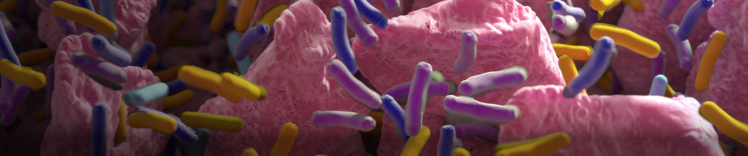
腸絨毛と細菌


微生物叢の構成要素
微生物叢(microbiome、マイクロバイオームともいう)は、ある特定の領域に生息する微生物(微生物相(microbiota))、その遺伝子、その微小環境(habitat(生息地))の総称です。細菌は、微生物叢の約 98% を占めています。1,2
微生物叢のサブセットには、ウイルス叢(virome)(ウイルス類)、真菌叢(mycobiome)(菌類)、古細菌叢(archaeome)(archaea(古細菌)、細菌に類似しているが別個の領域))などがあります。3微生物叢は、複雑な相互作用と相互に関連する代謝が存在する動的な環境です。4
ウイルス叢には、微生物叢内の細菌(バクテリオファージ)に感染するウイルス、環境内の宿主細胞に感染するウイルスが含まれます。5,6バクテリオファージは、ウイルス叢の大部分を占めています。5,6今までウイルス叢の研究は限られていましたが 6,7、慢性腸疾患または急性下痢症の犬において糞便内ウイルス叢の変化が特定されました。5,6同様に、ペットの微生物叢について発表されている研究論文の数は、現時点でも限られています。8
腸内微生物叢に存在する遺伝子の数は、宿主に存在する遺伝子の数をはるかに超えています。ヒトにおける微生物の遺伝子の数は、100 倍以上多いと推定されています。9
仔猫の腸内微生物叢をメタゲノム解析したところ、特定された一意の遺伝子の数は、猫のゲノムで特定されたオープンリーディングフレームの数よりも約 108 倍多かったことが示されました。10
腸内微生物叢中の存在比が最も高い細菌はファーミキューテス門(Firmicutes)であり、次いでバクテロイデス門(Bacteroides)およびフソバクテリウム門(Fusobacteria)、そしてプロテオバクテリア門(Proteobacteria)、アクチノバクテリア門(Actinobacteria)の順に続きます。11
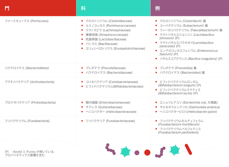
プロテオバクテリア門は、腸内微生物叢の中で最も多様な門であり、エシェリヒアコリ(Escherichia coli、大腸菌)、クレブシエラ(Klebsiella)、サルモネラ(Salmonella)、カンピロバクター(Campylobacter)など、多数の日和見病原体に加え、腸内ホメオスタシスに不可欠な役割を果たす細菌も含まれています。12猫の糞便中微生物叢は、犬より多様性が高い可能性があります。12
微生物叢フォーラムの他の領域を見る


詳しく知る
- Barko, P.C., McMichael, M.A., Swanson, K.S., Williams, D.A. (2018). The gastrointestinal microbiome: a review. Journal of Veterinary Internal Medicine, 32, 9–25. doi: 10.1111/jvim.14875
- Marchesi, J. R. & Ravel, J. (2015). The vocabulary of microbiome research: a proposal. Microbiome, 3, 31. doi: 10.1186/s40168-015-0095-5
- Kim, J. Y., Whon, T. W., Lim, M. Y., Kim, Y. B., Kim, N., Kwo, M.-S.,…Na, Y.-D. (2020). The human gut archaeome: identification of diverse haloarchaea in Korean subjects. Microbiome, 8, 114. doi: 10.1186/s40168-020-00894-x
- Seth, E. C., & Taga, M. E. (2014). Nutrient cross-feeding in the microbial world. Frontiers in Microbiology, 5, 350. doi: 10.3389/fmicb.2014.00350
- Moreno, P. S., Wagner, J., Mansfield, C. S., Stevens, M., Gilkerson, J. R., & Kirkwood, C. D. (2017). Characterization of the canine faecal virome in healthy dogs and dogs with acute diarrhoea using shotgun metagenomics. PLoS ONE, 12(6), e0178433. doi: 10.1371/journal.pone.0178433
- Moreno, P. S., Wagner, J., Kirkwood, C. D., Gilkerson, J. R., & Mansfield, C. S. (2018). Characterization of the fecal virome in dogs with chronic enteropathy. Veterinary Microbiology, 221, 38–43. doi: 10.1016/j.vetmic.2018.05.020
- Zhang, W., Li, L., Deng, X., Kapusinszky, B., Pesavento, P. A., Delwart, E. (2014). Faecal virome of cats in an animal shelter. Journal of General Virology, 95, 2553–2564. doi: 10.1099/vir.0.069674-0
- Foster, M. L., Dowd, S. E., Stephenson, C., Steiner, J. M., & Suchodolski, J. S. (2013). Characterization of the fungal microbiome (mycobiome) in fecal samples from dogs. Veterinary Medicine International, 2013, 658373. doi: 10.1155/2013/658373
- Richards, P., Thornberry, N. A., & Pinto, S. (2021).The gut-brain axis: Identification of new therapeutic approaches for Type 2 diabetes, obesity, and related disorders. Molecular Metabolism, E pub ahead of print. doi: 10.1016/j.molmet.2021.101175
- Deusch, O., O’Flynn, C., Colyer, A., Morris, P., Allaway, D., Jones, P. G., & Swanson, K. S. (2014). Deep Illumina-based shotgun sequencing reveals dietary effects on the structure and function of the fecal microbiome of growing kittens. PLoS ONE, 9(7), e101021. doi: 10.1371/ journal.pone.0101021
- Suchodolski, J. S. (2011). The intestinal microbiota of dogs and cats: A bigger world than we thought. Veterinary Clinics of North America Small Animal Practice, 41, 261–272. doi: 10.1016/j.cvsm.2010.12.006
- Belas, A., Marques, C., & Pomba, C. (2020).The gut microbiome and antimicrobial resistance in companion animals.In Duarte, A. & Lopes da Costa, L. (Eds.), Advances in Animal Health, Medicine and Production (1st ed.), pp. 233–245.Springer International Publishing