INTESTINAL VILLI WITH BACTERIA
Microbiomes Beyond the Gut
Although the gut microbiome has to date garnered the majority of attention, numerous microbiomes exist on and within the body.1
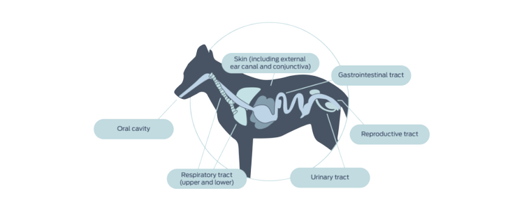
Microbiomes are present anywhere in the body where microbes reside, including the oral cavity, conjunctiva, external ear, skin, upper and lower respiratory tract, reproductive tract and urinary tract. Body sites differ dramatically in their microbial composition based on the physical and chemical properties of the site, including pH, topography and moisture level.2
Microbiome research in humans is rapidly expanding as investigation into non-gut microbiomes accelerates.
Investigation of non-gut microbiomes in dogs and cats lags behind those of humans, presenting additional opportunities to influence host health.


The oral microbiome
Despite its physical and functional connection to the gut, the oral microbiome of dogs and cats is a unique microbiome with about 50-100 million bacteria representing approximately 200 species.2-4 Studies to date have shown the oral microbiome of dogs and cats to be comprised of the same dominant phyla, but in different abundances between the species.3,5,6,7 In addition, the microbial population varies with location within the oral cavity.8
The oral microbiome appears to be highly conserved across dogs, with no significant differences between dog breeds.3 In contrast, significant differences in diversity and relative abundance of the oral microbiome were observed between several cat breeds as well as between cats with outdoor exposure and indoor-only cats.9
Alterations in the oral microbiome have been reported associated with birth method (c-section vs vaginal);5 diet format (wet vs dry, as well as changing diet from nursing to commercial food) in cats;5,10 feeding plaque-reducing dental chews to dogs;11 the administration of oral probiotics;12 dental prophylaxis;13 and oral disease (e.g., periodontitis, gingivitis, gingivostomatitis).14-18 However, whether the microbiome alterations precede and predispose to disease, or whether the alterations represent microbial population shifts in response to an altered environment, requires further investigation.3,19,20
Despite documented alterations, the oral microbiome is resilient.3,13
The skin microbiome
The skin microbiomes of dogs and cats are dominated by similar bacterial phyla, but are more diverse than the human skin microbiome.21 As observed in the oral microbiome, the skin microbiome of dogs and cats share the same predominant phyla but in different abundance.21 In both species, haired sites showed a higher number of microbes (richness) than mucosal and mucocutaneous junctions.21 The overall role of the skin microbiome in health and disease is poorly understood,22 but the skin’s role as a primary barrier and its close association with the immune system suggest it is a key player in host health.
The majority of research on the canine and feline skin microbiome has focused on the comparison of healthy and allergic or atopic dogs and cats.
Not surprisingly, research has documented significant differences in the skin microbiome of healthy and allergic cats as well as healthy and allergic or atopic dogs, with reduced diversity and richness associated with allergic and atopic disorders.21,23-26
Explore other areas of the Microbiome Forum
Find out more
- Yang, J. (2012, July 16). The Human Microbiome Project: Extending the definition of what constitutes a human. National Humane Genome Research Institute Genome Advance of the Month. https://www.genome.gov/27549400/the-human-microbiome-project-extending-the-definition-of-what-constitutes-a-human
- Koidl, L., & Untersmayr, E. (2021). The clinical implications of the microbiome in the development of allergy diseases. Expert Review of Clinical Immunology, 17, 115—126. doi:10.1080/1744666X.2021.1874353
- Bell, S. E., Nash, A. K., Zanghi, B. M., Otto, C. M., & Perry, E. B. (2020). Assessment of the stability of the canine oral microbiota a er probiotic administration in healthy dogs over time. Frontiers in Veterinary Science, 7, 616. doi:10.3389/fvets.2020.00616
- Dewhirst, F. E., Klein, E. A., Bennett, M.-L., Cro , J. M., Harris, S. J., & Marshall-Jones, Z. V. (2015). The feline oral microbiome: A provisional 16S rRNA gene based taxonomy with full-length reference sequences. Veterinary Microbiology, 175, 294—303. doi:10.1016/j.vetmic.2014.11.019
- Spears, J. K. (2017, May 4-6) Development of the oral microbiome in kittens. Proceedings of the Companion Animal Nutrition Summit, Vancouver, B. C., Canada, 73–81.
- Dewhirst, F. E., Klein, E. A., Thompson, E. C., Blanton, J. M., Chen, T., Milella, L.,…Marshall-Jones, Z. V. (2012). The canine oral microbiome. PLoS ONE, 7(4), e36067. doi:10.1371/journal.pone.0036067
- Sturgeon, A., Pinder, S. L., Costa, M. C., Weese, J. S. (2014). Characterization of the oral microbiota of healthy cats using next-generation sequencing. The Veterinary Journal, 201, 223–229. doi:10.1016/j.tvjl.2014.01.024
- Ruparelli, A., Inui, T., Staunton, R., Wallis, C., Deusch, O., & Holcombe, L. J. (2020). The canine oral microbiome: variation in bacterial population across different niches. BMC Microbiology, 20, 42. doi:10.1186/s12866-020-1704-3
- Older, C. E., Diesel, A. B., Lawhon, S. D., Queiroz, C. R. R., Henker, L. C., & Hoffmann, A. R. (2019). The feline cutaneous and oral microbiota are influenced by breed and environment. PLoS ONE, 14(7), e0220463. doi:10.1371/journal.pone.0220463
- Adler, C. J., Malik, Browne, G. V., & Norris, J. M. (2016). Diet may influence the oral microbiome composition in cats. Microbiome, 4, 23. doi:10.1186/s40168-016-0169-y
- Oba, P. M., Carroll, M., Alexander, C., Lye, L., Somrak, A., Keating, S.,…Swanson, K. S. (2020). Oral microbiota populations of adult dogs consuming dental chews demonstrated to reduce dental plaque and calculus. Journal of Animal Science, 98(Suppl 4), 61.
- Mäkinen, V.-M., Mäyrä, A., & Munukka, E. (2019). Improving the health of teeth in cats and dogs with live probiotic bacteria. Journal of Cosmetics, Dermatological Sciences and Applications, 9, 275–283. doi:10.4236/jcdsa.2019.94024
- Flancman, R., Singh, A., & Weese, J. S. (2018). Evaluation of the impact of dental prophylaxis on the oral microbiota of dogs. PLoS ONE, 13(6), e0199676. doi:10.1371/journal.pone.0199676
- Dolieslager, S. M. J., Bennett, D., Johnston, N., & Riggio, M. P. (2013). Novel bacterial phylotypes associated with the healthy feline oral cavity and feline chronic gingivostomatitis. Research in Veterinary Science, 94, 428–432. doi:10.1016/j.rvsc.2012.11.003
- Davis, E. M. (2016). Gene sequence analysis of the healthy oral microbiome in humans and companion animals. Journal of Veterinary Dentistry, 33(2), 97–107. doi: 10.1177/0909765416657239
- Nakanishi, H., Furuya, M., Soma, T., Hayashiuchi, Y., Yoshicuhi, R., Matsubayashi, M.,…Sasai, K. (2019). Prevalence of microorganisms associated with feline gingivostomatitis. Journal of Feline Medicine and Surgery, 21(2), 103–108. doi:10.1177/1098612X18761274
- Harris, S., Cro , J., O’Flynn, C., Deusch, O., Colyer, A., Allsopp, J.,…Davis, I. J. (2015). A pyrosequencing investigation of differences in the feline subgingival microbiota in health, gingivitis and mild periodontitis. PLoS ONE, 10(11), e0136986. doi:10.1371/journal.pone.0136986
- Older, C. E., de Oliveira Sampaio Gomes, M., Hoffmann, A. R., Policano, M. D., Cruz dos Reis, C. A., Carregaro, A. B.,…Carregaro, V. M. L. (2020). Influence of the FIV status and chronic gingivitis on feline oral microbiota. Pathogens, 9, 383. doi:10.3390/pathogens9050383
- Davis, I. J., Wallis, C., Deusch, O., Colyer, A., Milella, L., Loman, N., Harris, S. (2013). A cross-sectional survey of bacterial species in plaque from client owned dogs with healthy gingiva, gingivitis or mild periodontitis. PLoS ONE, 8(12), e83158. doi:10.1371/journal.pone.0083158
- Holcombe, L. J., Patel, N., Colyer, A., Deusch, O., O’Flynn, C., & Harris, S. (2014). Early canine plaque biofilms: Characterization of key bacterial interactions involved in initial colonization of enamel. PLoS ONE, 9(12), e113744. doi:10.1371/journal.pone.0113744
- Older, C. E., Diesel, A., Patterson, A. P., Meason-Smith, C., Johnson, T. J., Mansell, J.,…Rodrigues Hoffmann, A. (2017). The feline skin microbiota: The bacteria inhabiting the skin of healthy and allergic cats. PLoS ONE, 12(6), e0178555. doi:10.1371/journal.pone.0178555
- Weese, J. S. (2013). The canine and feline skin microbiome in health and disease. Veterinary Dermatology, 24, 173–e31. doi:10.1111/j.1365-3164.2012.01076.x
- Fazakerley, J., Nuttall, T., Sales, D., Schmidt, V., Carter, S. D., Hart, C. A., & McEwan, N. A. (2009). Staphylococcal colonization of mucosal and lesional skin sites in atopic and healthy dogs. Veterinary Dermatology, 20(3), 179–184. doi:10.1111/j.1365-3164.2009.00745.x
- Furiani, N., Scarampella, F., Martino, P. A., Panzini, I., Fabbri, E., & Ordeix, L. (2011). Evaluation of the bacterial microflora of the conjunctival sac of healthy dogs and dogs with atopic dermatitis. Veterinary Dermatology, 22(6), 490–496. doi:10.1111/j.1365-1365-3164.2011.00979.x
- Rodrigues Hoffmann, A., Patterson, A. P., Diesel, A., Lawhon, S. D., Ly, H. J., Elkins Stephenson, C.,…Suchodolski, J. S. (2014). The skin microbiome in healthy and allergic dogs. PLoS One, 9(1), e83197. doi:10.1371/journal.pone.0083197
- Bradley, C. W., Morris, D. O., Rankin, S. C., Chain, C. L., Misic, A. M., Houser, T.,…Grice, E. A. (2016). Longitudinal evaluation of the skin microbiome and association with microenvironment and treatment in canine atopic dermatitis. Journal of Investigative Dermatology, 13(6), 1182–1190. doi:10.1016/j.jid.2016.01.023

